[for you TL/DR people: MRI was good next one in two months]
This has gotten so mundane that I don’t even post about it anymore. Life just plugs along with the daily inconveniences of wearing sticky, itchy, annoying pads on my head. I am sure they will be even worse when it gets hot in summer. I had a great visit with Jessica Morris in NYC. She is a few months ahead of me and is an Optune wearer. She had problems with the pads overheating in the summer last year. So another thing to look forward to.
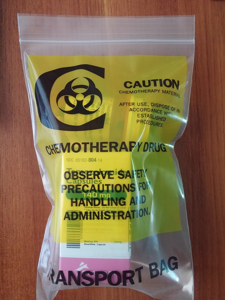
My Aunt Chemo still comes to visit for 5 days every month and she love to bring along the constipation just to let me know she loves me. Jessie and I have gotten better at dealing with this little gem and it has gotten better each round of Chemo.
So onto the good stuff. The MRI went fine – soon I will be able to sleep in that clicking, clacking, booop booop booop tube. There are two machines they seem to put the brain people in. The one I had today must be a slightly different model since the tests sound and feel different. The technician was new and she asked if I get these once a year – HA! I counted all of the times while I was in there and I am pretty sure I am up to 12 now, 11 of those at OHSU, 1 at Providence when they found the tumor. I really don’t remember that one much since I was so drugged up from the vertigo.
Today was the first time I actually got to see my scans the same day as my scan. There are two types of images they use to “read” MRI’s. The contrast is what they use to see if tumor is regrowing. The good news there is it does not look like it. Plus there are these two open areas called the ventricles. that run down the middle lower part of the brain. My Doctor told me when a tumor starts growing it pushes good brain mass aside and that often squashes into the ventricle. Mine looks great so it does not look like anything new is growing.
The second scan they look at does not have contrast and is called the FLAIR. Without getting super deep into the weeds it looks at the substance of the brain based on how it reacts to the magnetic field created by the MRI. Fat will look different from gray matter vs fluid in the brain etc. I did have one area that showed up quite large on the FLAIR. They think that may be from the radiation treatment. They referred to it as Gliosis. I will talk to my radiation oncologist about it tomorrow.
So after all that, they are happy with the scan as far as tumor progression, cautious about the potential radiation damage (I am going to blame that for my CRS syndrome [Can’t Remember Shit]), and will have me back in two months this time. Personally I prefer the two month schedule. I want to catch ANYTHING early when it comes to this thing growing back.
Will update tomorrow with my “2nd opinion”


